
 العربية
 فارسی
فارسی English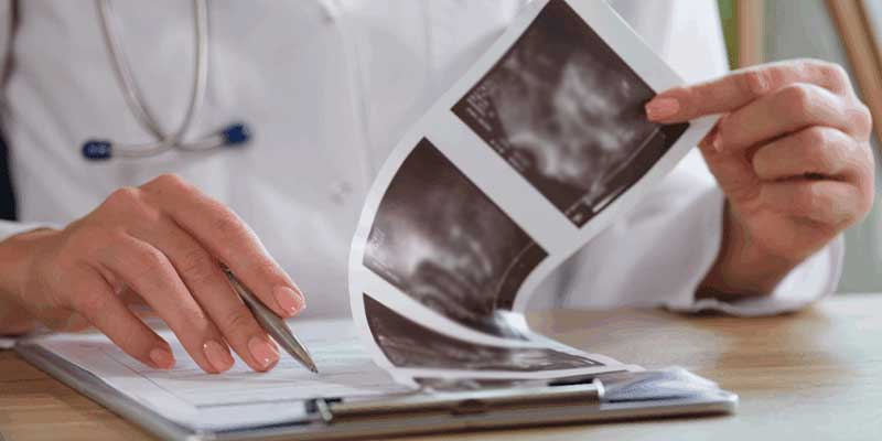
Table of Contents
What is a thyroid ultrasound report?
Thyroid ultrasound is a common imaging method used to examine the thyroid gland. Following the procedure, the results are presented in a medical report. This report typically details the size and shape of the thyroid, the condition of its tissue, the presence or absence of nodules (along with their specific characteristics), and other potential findings. However, the use of specialized medical terminology makes interpreting these reports difficult for many people. Below, we review the key terms and findings found in thyroid ultrasound reports to help you better understand what each section means and which findings might require further follow-up.
The importance of interpreting thyroid ultrasound results
Many people feel confused or anxious when they receive their thyroid ultrasound report due to unfamiliar medical terms. Words such as “hypoechoic,” “calcifications,” or “TI-RADS score” can seem alarming at first glance—especially if you do not know what they mean. The good news is that most thyroid ultrasound findings are not dangerous, and the majority of thyroid nodules are benign. Familiarizing yourself with the terms used in the report allows you to better understand your results before seeing a doctor and approach the process with less anxiety. Of course, the precise interpretation of the report and decisions regarding the need for follow-up or treatment must be made by a physician.
What does a thyroid ultrasound show?
Thyroid ultrasound creates images of the thyroid gland using sound waves, without the use of radiation. For this reason, it is a safe and common method for examining thyroid masses, monitoring changes and growth, or taking a closer look at findings observed via other imaging techniques. Reading a thyroid ultrasound report can be challenging if you are unfamiliar with imaging terminology. The report documents various details regarding the condition of the thyroid and any nodules or masses present.
Most thyroid ultrasound reports include the following information:
• Nodule location: Specifies whether the nodule is in the right lobe, left lobe, or the central part of the thyroid (isthmus).
• Nodule size: Usually recorded in three dimensions (in centimeters).
• Nodule composition: Indicates whether the nodule is solid, cystic, or a combination of both.
• Echogenicity: Shows whether the nodule appears lighter or darker compared to the surrounding normal thyroid tissue.
• Nodule margins and shape: Describes the appearance of the nodule’s edges and its overall shape.
• Calcification: Notes the presence of calcium deposits or calcified spots within the nodule.
Other findings that may be mentioned in a thyroid ultrasound report include:
• Cysts
• Goiter
• Thyroiditis
• Enlarged lymph nodes
These details are not merely supplementary information; by evaluating these specific characteristics, the physician assesses the level of concern regarding a nodule and determines whether follow-up, further investigation, or a biopsy is required. The report may also include a TI-RADS score. This system categorizes the nodule’s risk level based on its various characteristics, assisting the physician in deciding on the appropriate course of action, such as monitoring or biopsy. Therefore, to properly interpret a thyroid ultrasound report, it is best to consider these terms collectively rather than viewing a single word as an indicator of disease or cancer.
What is TI-RADS in a thyroid ultrasound report?
TI-RADS stands for “Thyroid Imaging Reporting and Data System.” This system was developed by the American College of Radiology (ACR) to provide a standardized and more understandable structure for thyroid ultrasound reports. Instead of relying solely on general descriptions to characterize a nodule, specific features are assigned points, and the total score determines the TI-RADS category. Simply put, each feature observed during the ultrasound is assigned a specific point value. The total score classifies the nodule into one of the categories ranging from TR1 to TR5. A higher category indicates more suspicious nodule features, potentially requiring closer monitoring or a biopsy.
The meanings of the TI-RADS categories are outlined in the table below.
Category Score Meaning Typical Action
TR1 0 Benign appearance Biopsy usually not required
TR2 2 No suspicious features Biopsy usually not required
TR3 3 Mildly suspicious Follow-up usually starting at 1.5 cm; biopsy usually starting at 2.5 cm
TR4 4–6 Moderately suspicious Follow-up usually starting at 1 cm; biopsy usually starting at 1.5 cm
TR5 7 or higher Highly suspicious Follow-up usually starting at 0.5 cm; Biopsy is typically considered for nodules 1 cm or larger.
Therefore, a high TI-RADS category does not automatically indicate thyroid cancer. For instance, a 2.8 cm nodule might fall into the TR3 category, yet a biopsy might be considered due to its size. Conversely, a smaller nodule in the TR5 category might require further evaluation due to its suspicious shape and textural characteristics.
Note: These figures pertain to the ACR TI-RADS system; thresholds for follow-up or biopsy may vary depending on the individual’s circumstances and the physician’s assessment.
Common Terms in Thyroid Ultrasound
Terms and words that patients commonly encounter in thyroid ultrasound reports include the following:
Thyroid Size & Shape
The ultrasound report assesses the overall size and appearance of the thyroid gland. A normal thyroid typically exhibits good symmetry and smooth, well-defined margins. Thyroid enlargement (goiter) may be associated with factors such as iodine deficiency, autoimmune thyroid diseases, or hormonal changes. Additionally, asymmetry or irregular thyroid margins are sometimes associated with inflammation, nodules, or structural changes.
Heterogeneous Thyroid Texture
A heterogeneous texture means the appearance of the thyroid tissue on the ultrasound is not uniform. This condition is observed in cases of thyroid inflammation (thyroiditis) or autoimmune diseases such as Hashimoto’s and Graves’ disease.
Thyroid Cyst
Cysts are fluid-filled lesions. Most thyroid cysts are benign and generally do not pose a serious risk; however, depending on their size and characteristics, a physician may recommend periodic follow-up.
Nodule
A thyroid nodule refers to an abnormal area or mass within the thyroid gland tissue that may appear as a protrusion on the ultrasound. Nodules are very common, and the majority are benign; however, some require further evaluation or monitoring. During the ultrasound, the nodule is assessed based on characteristics such as size, composition, echogenicity, shape, margins, and the presence of calcification. Taken together, these characteristics help determine the risk level of a nodule and guide decisions regarding follow-up or biopsy.
“Solid” or “Mixed”
Ultrasound reports indicate whether a nodule is entirely solid, cystic, or a mixture of solid and fluid components. This characteristic is one of the criteria considered in the TI-RADS system for nodule assessment. Solid nodules consist primarily of tissue, containing little to no fluid. In the TI-RADS system, these nodules typically receive higher scores than simple cysts. However, a solid nodule is not necessarily cancerous; many solid thyroid nodules are benign.
“Hypoechoic” (Low Echogenicity)
This term means the nodule appears darker than the surrounding thyroid tissue. Hypoechoic nodules may receive higher scores in the TI-RADS system, as this feature can be associated with an increased risk of malignancy. Nevertheless, echogenicity is just one of the features considered during assessment and cannot determine the risk level on its own.
“Calcifications”
Calcifications refer to tiny calcium deposits within the nodule. Not all types of calcification carry the same significance, and certain patterns may indicate a higher likelihood of malignancy. For instance, tiny, dot-like echogenic foci (punctate echogenic foci) may increase the risk of malignancy, whereas large, coarse calcifications are often observed in long-standing benign nodules. “Irregular margin” or “Taller-than-wide”
These are two characteristics radiologists consider when assessing the likelihood of a nodule being malignant. If a nodule’s height exceeds its width, or if its margins appear indistinct and irregular, it may receive a higher score in the TI-RADS system, as these features are less common in benign nodules.
Vascularity
Vascularity refers to blood flow within the nodule. Using Doppler ultrasound, a physician can check for the presence of blood vessels inside the nodule. Increased internal blood flow sometimes indicates heightened activity in that area; however, observing this finding in isolation does not necessarily mean the nodule is dangerous.
If your report mentions several of these features, do not interpret a single term in isolation. The TI-RADS score is determined based on the nodule’s overall set of characteristics, not just one specific phrase. Therefore, it is best to review the report with your physician and prepare any questions you may have about the results in advance.
Actions following ultrasound result interpretation
Based on the ultrasound findings, your physician might recommend one of the following:
• No further action: If the findings are entirely benign and no risk factors are present.
• Repeat ultrasound: If the nodule needs to be monitored at specific intervals to ensure there are no changes.
• Fine-Needle Aspiration (FNA): If the nodule is large, growing, or displays suspicious features on the ultrasound.
• Blood tests: To accurately assess thyroid hormone levels (such as TSH, T3, and T4). • Initiating treatment: If the physician suspects thyroid disease (such as hypothyroidism or hyperthyroidism), they will begin pharmacological treatment.
When are follow-up or biopsy considered?
Most thyroid ultrasound reports include a section at the end labeled “Conclusion” or “Impression,” where the TI-RADS classification and nodule size are listed together. This section is usually the most important part of the report for determining the next steps.
In general:
In general:
• Small TR1 or TR2 nodules usually do not require a biopsy.
• TR3 nodules are often monitored via follow-up ultrasound to check for growth or changes before considering a biopsy.
• TR4 and TR5 nodules are more likely to require fine-needle aspiration (FNA) if they reach the size thresholds specified in the ACR system.
• Nodule growth over time is also significant, even if no concerning features were observed on the initial ultrasound.
• If a biopsy is required, ultrasound guidance is used to accurately position the needle at the target site, allowing the physician to sample the specific area of concern.
• If you encounter a phrase like “recommend follow-up ultrasound in 12 months” in your report, it usually means a re-examination is suggested to monitor the nodule’s status and ensure stability—not that something important was overlooked during the previous scan.
• The phrase “recommend FNA” (fine-needle aspiration) indicates that the radiologist has determined that analyzing the tissue cells could provide more precise information regarding the nature of the nodule.
The Bottom Line
A thyroid ultrasound report is not a definitive diagnosis; rather, it is a summary of the findings observed by the radiologist during the examination of the thyroid and its nodules. Each term in the report describes a specific characteristic of the nodule, and these features collectively help assess the level of risk. Ultimately, your physician will decide on the next steps by considering the ultrasound results alongside your overall clinical situation.



No comment